Tomography, Free Full-Text
Tomography, Free Full-Text
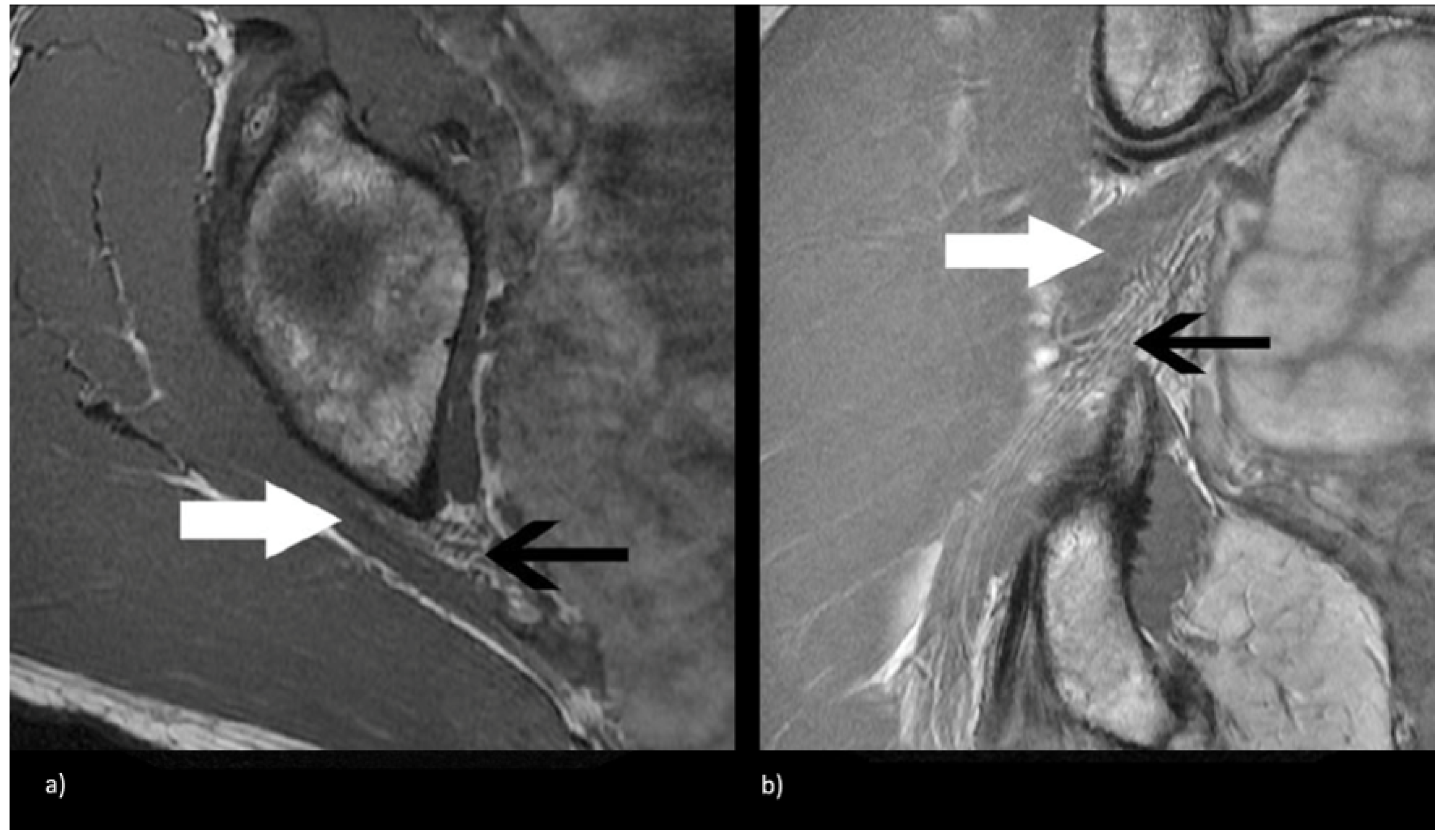
Objective: To assess the prevalence and clinical implications of variant sciatic nerve anatomy in relation to the piriformis muscle on magnetic resonance neurography (MRN), in patients with lumbosacral neuropathic symptoms. Materials and Methods: In this retrospective single-center study, 254 sciatic nerves, from 127 patients with clinical and imaging findings compatible with extra-spinal sciatica on MRN between 2003 and 2013, were evaluated for the presence and type of variant sciatic nerves, split sciatic nerve, abnormal T2-signal hyperintensity, asymmetric piriformis size and increased nerve caliber, and summarized using descriptive statistics. Two-tailed chi-square tests were performed to compare the anatomical variant type and clinical symptoms between imaging and clinical characteristics. Results: Sixty-four variant sciatic nerves were identified with an equal number of right and left variants. Bilateral variants were noted in 15 cases. Abnormal T2-signal hyperintensity was seen significantly more often in variant compared to conventional anatomy (40/64 vs. 82/190; p = 0.01). A sciatic nerve split was seen significantly more often in variant compared to conventional anatomy (56/64 vs. 20/190; p < 0.0001). Increased nerve caliber, abnormal T2-signal hyperintensity, and asymmetric piriformis size were significantly associated with the clinically symptomatic side compared to the asymptomatic side (98:2, 98:2, and 97:3, respectively; p < 0.0001 for all). Clinical symptoms were correlated with variant compared to conventional sciatic nerve anatomy (64% vs. 46%; p = 0.01). Conclusion: Variant sciatic nerve anatomy, in relation to the piriformis muscle, is frequently identified with MRN and is more likely to be associated with nerve signal changes and symptomatology.

Incidental Findings on Brain MRI in the General Population
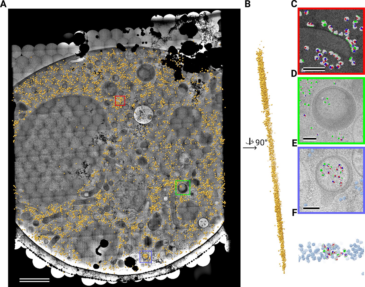
Defocus Corrected Large Area Cryo-EM (DeCo-LACE) for label-free detection of molecules across entire cell sections
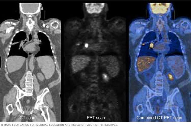
Positron emission tomography scan - Mayo Clinic

Multiclass semantic segmentation and quantification of traumatic brain injury lesions on head CT using deep learning: an algorithm development and multicentre validation study - The Lancet Digital Health

PET/CT in Brain Disorders

Optical Coherence Tomography and Eye Care
Imaging techniques for the diagnosis of soft tissue tumors

Dark-field computed tomography reaches the human scale

Computed Tomography


